
The Gold standard Intervention for GORD: Laparoscopic Nissen’s fundoplication
First ever Nissen’s fundoplication was done in 1936; from 1956 onward the operation found its place as an effective treatment for Gastro Oesophageal Reflux Disease.
Many more anti reflux Fundoplication procedures developed from 60’s to 80’s. In 90’s some endoscopic procedures were also developed and these have continued to evolve to date. The oldest and original operation of Nissen’s Fundoplication remains as the gold Standard with best short- and long-term results in the hands of expert surgeons.
With the advent of Laparoscopic Surgery in late 80’s, this operation became popular. The operation is truly minimal invasive with quick recovery and can offered to suitable patients as a day case procedure as well.
Surgery is done under general anaesthetic. The operation itself can be accomplished between 45 minutes to an hour.
Fine ports and instruments are used to perform surgery. In all five ports, which are small and narrow tubes with valves are placed in abdomen to fill some gas to distend the abdomen. This makes space for seeing the structures and carry on the operation.
Some of the key components of surgery are:
- A 10 mm camera port is placed in the middle of abdomen. Gas is filled in abdomen at a low pressure and laparoscopy (look around inside the abdomen) is performed.
A retractor is placed in abdomen to lift the liver from the front of the stomach, diaphragm and hiatus to make space for visualisaton and surgery.
Two other small 5-mm ports and one 10 mm port are placed in the upper abdomen.
- Fine instruments are used to identify hiatus and it is dissected free and separated from the oesophagus.
- Fundus is freed from its attachments to the spleen on the left side and diaphragm above.
- Further mobilisation of the esophagus and hiatus hernia is carried out high up in the chest to get adequate intra-abdominal length of esophagus (more than 6 cm).
- Lax hiatus is repaired (Cruroplasty) with non-absorbable 4-5 stiches. This brings it back to its normal size.
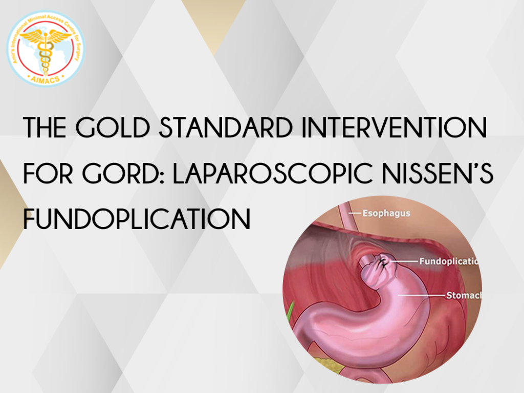
- A floppy 2 cm long 360̊ wrap done after assuring appropriate positioning of the fundus around the lower end of the esophagus with three non-absorbable stiches.
- Above the warp there is some intra-abdominal oesopghageal length without retraction.
- This completes the correction of normal anatomy by:
- Reducing the hiatus hernia and bringing the stomach back to abdomen
- Tightening of the Hiatus with permanent stitches to prevent formation of another hernia
- Having a decent length of Oesophagus in the abdomen.
- Creation of an angle (“of His”), between the fundus and the lower end of food pipe/ gullet.
11. Skin is closed with very fine stiches not visible to eye.
20. Sterile dressings are applied.
Postoperative Instructions:
Postoperative recovery is generally straight forward.
Patient is mobilised early on the day of surgery.
Patient is generally allowed to go home the day after the surgery after an overnight stay in the ward. Routine observation are carried out in the ward.
Patient can start drinking once comfortable and awake.
Patients are allowed free fluids post operatively building to sloppy/soft diet by the end of the day.
Regular pain killers and anti-sickness medication are given for 4- 5 days.
Dietician reviews the patient and advises on diet plan.
Patient is advised soft diet, building to normal diet over two weeks.
One can commence driving in 4-5 days after surgery.
One to Two weeks of leave from work will be required.
No heavy lifting for six weeks.
Follow up involves a review with operating surgeon in clinic 1 week and then 6 weeks after the surgery.