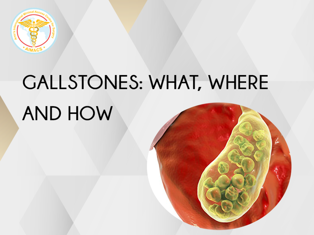
Gallstones: What, Where and How?

At what age one develops gallstones?
Common age group is 35- 55 years. However, one can have this as a newborn or even in 90s.
Symptoms of Gallstones:
Pain from gallstones generally occurs in the right upper abdomen and it radiates to the back between the shoulder blades. Sometimes the pain from gallstones can just be in upper abdomen without radiating to the back.
The pain is severe and females often describe this as worse than labour pain.
What causes pain ?
Pain from gallstones can be triggered by eating a fatty meal or drink. Fatty meal causes strong contraction of gall bladder on the stones. It’s like walking on a pebble beach without slippers. At times the stone may become impacted in the outlet (neck) of gall bladder.
Do all gallstones cause symptoms?
No, most gallstones remain asymptomatic and one may not be aware of their presence.
What are Gallstones?
Gallstones, medically known as cholelithiasis, are small or large stones present in the gall bladder. Gall stones can be very small or large, single or multiple. Mostly the gallstones have a smooth surface. The colour can range from pale whitish (cholesterol stones) to dark brown (Pigment stones)
What is Bile?
Bile is a liquid produced in the liver from 500 mls to one liter in a day. It exits the liver through the tubes (right and left hepatic ducts), most of it enters the small intestine directly, some enters the gall bladder where it is concentrated. Whenever signals get to gall bladder after eating food, gall bladder contracts and the bile from the gall bladder flows into the duodenum (small bowel).
Does bile digest fat?
No, it just helps top break it down into smaller particles like “dish washing liquids”, then it becomes easier for water to wash it away. The digestion of fat in the gut is carried out by enzymes from the gland pancreas.
Gall Bladder Anatomy:
Gall bladder is a small pear-shaped organ attached to the lower surface of liver. It capacity is only 50-70 mls. It is attached by a ting tube (Cystic duct) to the main tube (common bile duct) connecting the liver to the duodenum (small bowel).
Function (purpose) of Gall Bladder:
Gall bladder stones and concentrates a small amount to bile. The bile is continuously produced in the liver and it keeps trickling into the intestine. Between 500 mls to 100 mls of bile is produced by liver in 24 hours. The concentrated bile from the gall bladder is emptied into the duodenum (first 25 cm of small bowel) in response to the signals generated by gut after eating food; especially fatty food.
What causes gallstones formation?
There can me many reasons. Infection of gall bladder, infection of bile, poor contraction or obstruction of gall bladder and change in the constituents of the liquid bile are some of the common causes.
How the stones are formed in Gall Bladder?
Initially the infection, stasis or altered chemistry of bile causes some small deposits of bile or cholesterol pigments in the gall bladder, described as sand particles. Over the course of time more deposition on the top of these changes the small particles into small stones. Some can become very large in size over period of time. Some large stones can be of the size of and shape of an egg.
INCIDENCE/EPIDEMIOLOGY
Gallstones are a common medical condition all over the world. In United Kingdom alone about 80,000 to 100,000 gall bladder surgeries are performed yearly for gallstones. The actual number of people harboring stones in the general population is much higher.
Cholesterol and mixed stones are common in Europe and United States, whereas stone is more common in Asians and Africans.
RISK FACTORS
Immutable risk factors and associations:
Its more common in females over the age of 40.
Common in Caucasians.
Genetics and family
Modifiable Risk Factors:
Obesity
Metabolic syndromes
Pregnancy
High-fat diet
Diabetes
Cholesterol rich diet
Poor fiber diet
Certain blood disorders like sickle cell anemia or leukemia
Medications containing estrogen, such as oral contraceptives or HRT drugs
Rapid weight loss
Diseases like Liver cirrhosis and Crohn disease
Gallbladder stasis (due to spinal injury or drugs, like somatostatin),
Sedentary life
Gallstones presence can lead to numerous symptoms and complications when they are within the gallbladder. When they escape the gall bladder and enter the common bile duct then the complications are different and can be life threatening as well.
Symptoms and complications of Gallstones when inside the gallbladder:
These can cause vague symptoms as nausea, change in taste or mild fever.
Flatulent Dyspepsia
Commonly observed in obese, fatty women complaining of heartburn, abdominal discomfort, belching and fatty food intolerance.
Pain / Gallstone colic
Medically termed as biliary colic. Its Severe, colicky pain felt mostly in the upper abdomen. Pain can shoot to the back or between the shoulder blades often associated with vomiting, restlessness, and sweating. Strong pain killers, clinical assessment and sometimes admission in the hospital is required.
Dyskinesia of gallbladder (poorly contracting gallbladder):
Patients can have gall bladder pain even without gallstones. Specialist surgical opinion will help to solve this mystery; which can be debilitating for patients and can take a long time to diagnose.
Infection / Inflammation /
Acute Cholecystitis in medical terms occurs when a gallstone gets lodged in the neck of the gallbladder (Hartmann’s pouch), causing bile stasis leading to gallbladder inflammation. Cholecystitis is an acute condition and can cause severe pain, vomiting, nausea, and fever. Strong pain killers, clinical assessment, blood tests, antibiotics and admission in the hospital is required.
Acalculous Cholecystitis (Infection of Gall bladder without any stones):
This condition is noted in very sick patients and requires specialist input of an experienced surgeon. What may look like an inflamed gallbladder on scan be a gangrenous gall bladder.
Mucocele
It occurs when a stone is completely blocking the gall bladder outlet blocking the cystic duct or the Hartman’s pouch, but there is no infection. This leads to a distension of the gall bladder and pain. One notices “white bile” in the gall bladder at surgery. Surgery is required in this situation.
Empyema
It’s a completely blocked gall bladder like a mucocele with superadded infection as well. Its like an abscess, where simple antibiotics will not work. Emergency decompression or removal of gall bladder is required after appropriate assessment by a surgeon in a hospital.
Perforation of gall bladder
Perforation in abdominal cavity is a rare condition. Impacted stone, diabetes, or virus can precipitate an empyema causing gangrene of gallbladder and perforation.
High-grade fever, chills, and pain in the abdomen are common symptoms. Emergency treatment in a hospital with immediate surgery can be lifesaving in this situation.
Fistula formation:
In case on chronic cholecystitis a stone in gall bladder can erode into duodenum or large bowel, leading to Cholecystoduodenal fistula or a Cholecystocolonic fistula. This can happen silently and may come to attention while having tests done for any reason. Generally these can be left alone and observed it not giving any symptoms to the patient.
Mirizzi’s Syndrome:
A stone impacted in the Hartman’s pouch or the cystic duct if not causing a full blockage of the gallbladder outlet can keep getting bigger and can fistulate (erode) into the common bile duct.This can lead to pain, infection, jaundice or even sepsis and requires input from a specialist surgeon urgently. This should ideally be dealt with Laparoscopically and may also require an ERCP (Endoscopic Retrograde Cholangio Pancreaticography).
Small bowel Obstruction / Gallstone Ileus:
A large gallstone escaping the gall bladder due to Cholecystoduodenal fistula can get impacted in small bowel and cause a complete blockage requiring emergency surgery to relive the obstruction and remove the gallstone from small bowel.
Gallbladder Cancer:
The likelihood of cancer is not common, but chronic cholecystitis or cholelithiasis can pose an increased risk of gallbladder cancer. Gallbladder polyps and Porcelain gall bladder are also considered as risk factors for gallbladder cancer.
Gall Bladder cancer carries a poor prognosis.
B. Complications of Gallstones in the Common Bile Duct (CBD)
Though the complications are fewer than of the stones within the gallbladder but these are much more serious and potentially life threatening.
Obstructive Jaundice:
Obstruction to the bile outflow causes obstructive jaundice. Jaundice with yellow skin, yellow eyes, dark colored urine, clay-colored stools, and itching are the usual symptoms.
One need to see a surgeon to get the advice and treatment.
Generally, one requires ERCP and Laparoscopic Cholecystectomy.
Cholangitis:
It is the bacterial infection of common bile duct causing abdominal tenderness, chills, fever, and rigors. This is a very scary situation for the patient and requires urgent attention, admission in the hospital and antibiotic treatment with appropriate decompression of the system.
Gallstones Pancreatitis
There is no greater or worse complication of gallstones than acute pancreatitis or inflammation of pancreas due to a gallstone blocking the common opening of the common bile duct with the pancreatic duct.
Patient requires emergency admission in the hospital and appropriate care, investigations and treatment.
In mild pancreatitis one may get better in a day or two. In moderate cases one may lose the function of pancreas and become diabetic for life long. In some cases of severe pancreatitis, this may me life threatening.
Clinicians suggesting or patients considering wait and watch on their gallstones must consider this small but potentially life-threatening complications of gallstones in their mind.
GALLBLADDER ASSESSMENT
Gallstones infection (cholecystitis) or obstruction (empyema) presents with colicky pain in the upper abdomen with severe nausea, vomiting, and low-grade fevers initially.
Clinical Assessment
- Positive Murphy’s Sign (tenderness below right ribs)
- Boas’s Sign (Hypoaesthesia between shoulder blades)
- Upper abdominal guarding and rigidity
- Abdominal Tenderness
GALLBLADDER INVESTIGATIONS
Blood Tests:
Full blood count (white cell count is raised)
CRP is elevated
Liver Function tests
Amylase
Abdominal Ultrasound
It is a simple, painless, and noninvasive test, the same as pregnancy scans. Gel is applied to the abdomen, and a probe is used to bounce high-frequency sound waves off structures in the abdomen.
Ultrasound gives a clear view of the abdominal organs and is an excellent test for checking the gallbladder wall thickening and the gallstone diagnosis.
CT scan
It is done when ultrasound findings are not clear. It provides good views of the pancreas and helps with the diagnosis of pancreatitis. Its not the best test for stones in the gall bladder and less sensitive than an ultrasound.
Magnetic resonance cholangiopancreatography (MRCP)
An MRI scan provides valuable information in gallbladder diagnosis. Using magnetic waves, it provides high-resolution images of the biliary tree, ductal system, pancreas, and gallbladder. MRCP is required when stones are suspected in the common bile duct.
Endoscopic retrograde cholangiopancreatography (ERCP)
ERCP is an endoscopic procedure in which a flexible tube is inserted through the mouth, stomach, and into the small intestine. Then the doctor injects dye and with fluoroscopy can see the bile duct system. When stones are confirmed, then one can remove these by cutting the sphincter of Oddi (tip of common bile duct opening) and retrieving all the stones. Sometimes a stent (fine plastic tube) is left behind to maintain the flow of bile. Laparoscopic Cholecystectomy can be done in the same setting to avoid another anaesthetic.
Endoscopic ultrasound
A small, flexible tube with an ultrasound probe on end is inserted through the mouth to the intestines. Choledocholithiasis and gallstone pancreatitis can be diagnosed through endoscopic ultrasound; though rarely.
HIDA scan (cholescintigraphy)
Its used to check the function of the gall bladder in case of. HIDA Scan is a nuclear test in which a radioactive dye is injected intravenously and is secreted into the bile. The natural path of bile is followed, and if the scan shows the failure of bile ejection from the gallbladder.
Abdominal X-ray
Not usually used for gall stones. Abdominal X-rays can show gallstones, but does not give a definitive diagnosis. Sometimes it can confirm gallstone Ileus; still a CT scan will be required to confirm the diagnosis.
SELF-TREATMENT
Gallstone pain is acute and sharp in nature. Self-treatment options are limited. Some are:
Painkillers
Over the counter, painkillers can provide temporary relief in gallstone pain. However, in the case of acute cholecystitis, OTC painkillers will not be very effective and one will need to see a doctor.
Heat Therapy
Applying heat relaxes muscles, relieves pain, and is soothing. A heated compress releases tension due to bile build-up and provides relief. This is should only be used temporarily while seeking medical advice.
Peppermint Tea
Menthol promotes pain relief. Peppermint tea does not provide immediate relief, but regular drinking can reduce gallbladder flare-ups and pain in only some cases.
SEEKING MEDICAL ADVICE
Gallstone pain can wax and wane. It can remain asymptomatic for long periods of time but can always flare up unexpectedly. Seeking medical opinion on time is beneficial in terms of health, time, resources and avoiding complications.
If you are experiencing any of these symptoms, then see a doctor immediately.
- Nausea and vomiting
- Prolonged abdominal pain lasting more than 4 hours
- Fever
- Chills and rigors
- Yellowish tinge in the white of the eyes or skin
- Clay-colored stools
- Fatty food intolerance
- Dark-colored urine
MANAGEMENT
The following options are viable to avoid gallstones in the first instance.
- Conservative Lifestyle sedentary life is the root cause of many health issues. Obesity and high cholesterol are some of the factors of gallstones. Regular exercise and a balanced diet maintain weight and cholesterol levels.
Gallstone risk can be minimized by keeping a few points in mind.
- Avoid skipping meals. Try to maintain a routine of your meals for each day. Irregular meals and fasting can increase gallstone risk.
- Rapid weight loss can cause gallstone. Try and lose weight slowly.
- Eat high-fiber, low cholesterol foods. Vegetables, grains, and fruits make a balanced diet.
- Try to maintain a healthy weight.
Temporary measures:
Exercise and high-fiber, low-fat diet prevents gallstones may reduce the pain flare ups in gallstone patients. For gallstone patients, this is a temporary treatment option while waiting for a definitive medical intervention, which is necessary to avoid any complication.
- Medical Treatment
Medical Treatment was suitable obly for some small stones. Once popular in 1970’s it is not used anymore. Medication was used to dissolve stones. Medicine had to be used for may months or a few years. Soon after stopping the medication patients would get stone recurrence.
- Shock Wave Lithotripsy (SWL)
While useful in Kidney stone SWL or Laser treatments are not an option for stones in
the gallbladder.
- Olive oil remedies, mixtures and potions
These are not recommended and can be very dangerous if strong contraction of the gallbladder will expel the stone on the common bile duct, thus causing with potential complications alike acute pancreatitis, which could be life threatening.
- Surgical Treatment
- surgical removal is the gold standard treatment option for gallstones. This can be offered safely in experienced hands and prevents complications and the complete removal of gallstones.
- Operation in the form of Laparoscopic Cholecystectomy is the most commonly performed surgical procedure that involves four small cuts in the abdomen. A laparoscope (a camera with light source) is introduced in the abdomen. . The surgeon Introduces 3 fine instruments, not much thicker than chopsticks to separate the gallbladder from the adjoining organs and remove it carefully through one of the small incisions.
Laparoscopic Cholecystectomy is a minimally invasive procedure which is done as one night stay in the hospital. This can be performed in suitable patients as day case as well.
RISKS OF WAITING
- Acute gallbladder inflammation (cholecystitis)
- Bile duct stone (choledocholithiasis)
- Acute bacterial infection (cholangitis)
- Pancreatic infection (pancreatitis)
- Perforation of gall bladder
- Cholangitis (severe infection)
SURGICAL RISKS FACTORS
Laparoscopic Cholecystectomy is well established for more than 30 years. It is performed under full general anaesthetic. It takes a good surgeon and team about 15 – 25 minutes to perform a standard Laparoscopic Cholecystectomy.
Prior to this the operation of removal of gall bladder was done with long open cut just below the right ribcage. Hospital stay used to be 5-7 days after surgery and recovery would take weeks and months.
Risks of Cholecystectomy open or Laparoscopic are:
- Bleeding
- Damage to the neighboring organs such as liver or intestine
- Damage to common bile duct (very serious complication)
- Postoperative infection
- Postoperative pain
No surgery should be taken lightly. The above-mentioned risks all combined are quoted as less than 1%. The complications mentioned can be severe, may require a reoperation. The complication of damage to common bile duct is very serious indeed and much more common than generally quoted. In United Kingdom its incidence is 1:250, which unfortunately is unacceptably too high.
In experience hands of specialist upper Gastro Intestinal (Upper GI) surgeons the incidence of damage to common bile duct will lower than 1:1000.
In case of this rare occurrence in upper GI surgeon’s hands they ae able to reocgnise it and deal with this skillfully and in a timely fashion.
GOLD STANDARD TREATMENT FOR GALLSTONES
Currently, laparoscopic cholecystectomy is the gold standard for gallstone treatment. It has fewer complications, rapid recovery, less post-op pain, early mobilisation, return to driving, and early return to work and exercise.
It also provides quick and effective treatment for gallstones and prevents recurrence and complications.


Hi, this is a comment.
To get started with moderating, editing, and deleting comments, please visit the Comments screen in the dashboard.
Commenter avatars come from Gravatar.
Pingback: What are Gallstones? – Nissen’s Fundoplication